RADIOGRAPHS

Imaging technique that uses radiation to view the internal form of an object to evaluate your oral health. This can help dentist to identify problems, like cavities, tooth decay and impacted teeth.

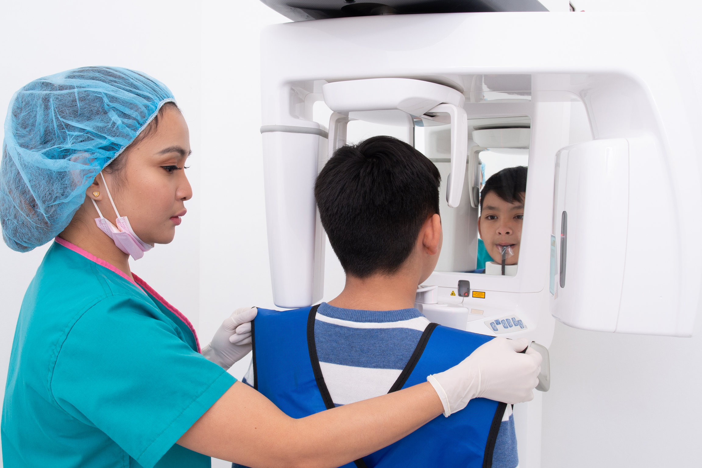
PANORAMIC

Uses a very small dose of radiation to capture the entire mouth in one image. It used to plan treatment for dentures, braces, extractions and implants.

CEPHALOMETRIC

A cephalometric x-ray is a diagnostic radiograph which shows us the skull in side view which is primarily used for orthodontic treatment planning.

PERIAPICAL

A periapical x-ray is a focused x-ray that shows the whole tooth from crown up to the apex. Periapical x-ray detects unusual changes in the root and surrounding bone structures.
BITEWING

Shows detail of the upper and lower teeth in one area of the mouth. Each bitewing shows a tooth from its crown (the exposed surface) to the level of the supporting bone. Bitewing X-rays detect decay between teeth and changes in the thickness of bone caused by gum disease. Bitewing X-rays can also help determine the proper fit of a crown.

TMOGRAM (TMJ)

Allows visualization of the articular tubercle, mandibular condyle and fosse and is thus useful to identify structural changes and displaced fractures, as well as assess excursion and joint spaces.

RADIOGRAPHS

Imaging technique that uses radiation to view the internal form of an object to evaluate your oral health. This can help dentist to identify problems, like cavities, tooth decay and impacted teeth.

Uses a very small dose of radiation to capture the entire mouth in one image. It used to plan treatment for dentures, braces, extractions and implants.

A cephalometric x-ray is a diagnostic radiograph which shows us the skull in side view which is primarily used for orthodontic treatment planning.

A periapical x-ray is a focused x-ray that shows the whole tooth from crown up to the apex. Periapical x-ray detects unusual changes in the root and surrounding bone structures.

Shows detail of the upper and lower teeth in one area of the mouth. Each bitewing shows a tooth from its crown (the exposed surface) to the level of the supporting bone. Bitewing X-rays detect decay between teeth and changes in the thickness of bone caused by gum disease. Bitewing X-rays can also help determine the proper fit of a crown.

Allows visualization of the articular tubercle, mandibular condyle and fosse and is thus useful to identify structural changes and displaced fractures, as well as assess excursion and joint spaces.